
Tomorrow Will Not Be Different
Dysthymia is a form of low-grade depression that never quite leaves. Let’s discuss why it seeps throughout every aspect of life and is so notoriously hard to treat.

Dysthymia, also known as persistent depressive disorder (PDD), is a long-term condition that lasts for at least two years in adults, or one year in children and teens. The symptoms are less severe than major depressive disorder (MDD) but equally debilitating. Its unifying features are low mood or sadness, fatigue, poor self-esteem, changes in appetite, difficulty concentrating, and persistent feelings of hopelessness. The difficulty of treating dysthymia lies in its chronic, self-reinforcing, and identity-entwined nature.
(IMPORTANT: this article is not medical advice, and has not been peer reviewed. You should consult your doctor if you have questions or feel unwell).

Dysthymia in two paragraphs
Unlike major depressive disorder, dysthymia becomes part of people’s personality over time. One of the reasons, and main differences with major depressive disorder, is its duration. Episodes of major depression generally last approximately 2 weeks, whereas dysthymia lasts for 2 years or more. Therefore, whereas major depressive disorder comes and goes, dysthymia lingers and seeps through every aspect of life. People with dysthymia often forget what normal life is like. The low mood becomes part of their concept of self. They often rationalize it by saying “this is just who I am.” This makes them less likely to seek help or to believe that improvement is possible. Consequently, even neutral events like a task or a an unexpected look from someone can be decoded negatively. Their mind starts to acquire habits of negative interpretation. “I will probably mess this up”, or “this is nice but it is not going to last anyway” are commonplace for the dysthymic mind. When these thoughts become automatic, they begin to shape perception and emotion, reinforcing negative feelings.
People with dysthymia often remain high-functioning, but simply because they “manage.” However, one cruel feature of dysthymia is the emotional flattening. Their brain learns to protect itself from sadness (which is a completely different emotion that we discuss in another article), but at the cost of lowering sensitivity and muting everything. As a consequence, success doesn’t feel exciting anymore, connections don’t feel warm, and even a good night sleep is not restorative. People with dysthymia often claim that it is like watching life instead of living it. Over time, the mind creates a distorting feedback loop where the external world begins to match the internal one. Eventually, people living with dysthymia choose less stimulating routines, withdraws socially, and avoid opportunities for reward. Life has less color and fewer emotional highs, and people stop expecting to feel any different. Dysthymia is normalized and becomes self-perpetuating.
Not just in the mind
The mind is the intangible realm of consciousness, thoughts, emotions, perceptions, and the sense of self. It is not located in a specific place but emerges from the dynamic patterns of brain activity. The brain, by contrast, is an actual organ. It is a tangible structure located in the head. It is made of neurons, glial cells and blood vessels. The brain is the body’s central control system that uses electrical and chemical signals. Therefore, you can see it on an MRI, dissect it with scalpels and study its molecules. Despite these fundamental differences, just like the mind the brain adapts to dysthymia to further worsen the condition. Because dysthymia is long lasting, the brain has time to rewrite how to handle it. It’s neurochemical baseline shifts. For example, the prefrontal cortex, especially the sectors supporting decision-making, cognitive control, and motivation show some of the most consistent changes, including altered activity and connectivity. This biases people’s attention toward negative information and to reduce top-down regulation of emotion. This means that the brain tends to focus more on negative experiences than on positive ones. The mind, then, becomes less effective at controlling emotional reactions, making it harder for people to regulate feelings rationally. It is as if the brain is a radio that has slowly drifted off station. Instead of giving music, it mostly catches static and the tuning knob is out of reach. Some studies indicate that neural circuits that are implicated in acute forms of depression are also affected in chronic forms such as dysthymia. For example, the amygdala, which underlies emotions, rewires so that it generally lowers its response to general stimuli, or increases its responses to negative cues. One experimental study manipulated norepinephrine signaling and found that boosting norepinephrine produced a negativity bias in the amygdala, in that the amygdala showed increased responses to negative stimuli and simultaneously decreased responses to positive stimuli (#here). The amygdala also interacts with dopaminergic systems, especially in salience, novelty, reward prediction. It has been well established that dysregulation of dopamine occurs in depression, for example in anhedonia, and influences brain circuits including the amygdala indirectly. Together, this negative bias makes the brain interpret neutral experiences as bad, filtering out anything that doesn’t fit the gloomy pattern.
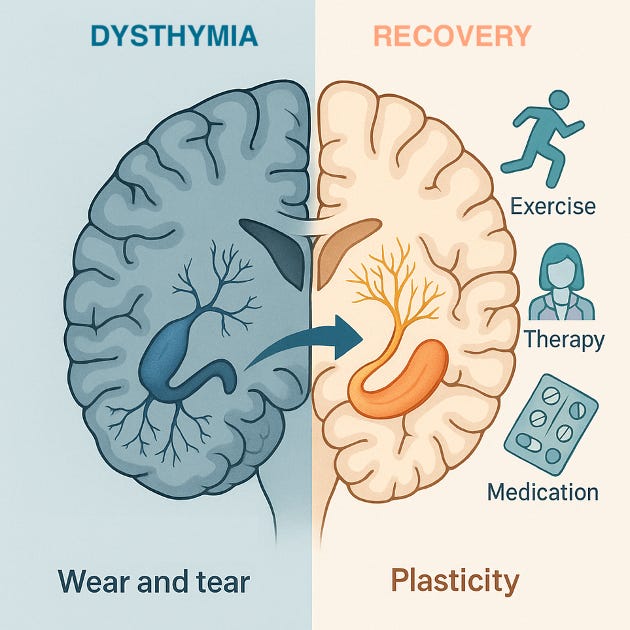
Another important brain region that becomes a bad actor is the hippocampus, because it starts to operate in a low-state. Multiple magnetic resonance imaging (MRI) studies have shown that people with dysthymia have a smaller hippocampal volume compared to healthy controls. This is similar to what has been observed in major depressive disorder, but in dysthymia the changes are more gradual and subtle. The hippocampus also helps contextualize emotional experiences and regulate the stress-response axis. In dysthymia, the impaired hippocampus fails to turn off the stress response properly, maintaining a chronic activation of stress hormones. In addition, chronic low serotonin and dopamine levels mean the reward system dulls, and things that used to bring pleasure barely register. Human PET studies and meta-analyses implicate changes in the serotonin 1A (5-HT1A) receptor in depression, consistent with serotonergic dysregulation that could reflect (or drive) a new “set-point” in mood regulation over time. Although findings vary, they converge on 5-HT1A alterations in cohorts of people with depression. This further contributes to a negative cognitive bias when the brain stops integrating new positive memories effectively, helping old negative emotions dominate. Functionally, this means things that should feel good barely move the needle, and people invest less and less effort to pursue rewarding activities (#here). Moreover, the long-term stress common to dysthymia leads to elevated glucocorticoid levels (think cortisol), which can damage or shrink hippocampal neurons and suppress restorative neurogenesis. Long-term exposure to elevated glucocorticoids decreases serotonin signaling, reduces neurotrophic support, particularly brain-derived neurotrophic factor (BDNF), and gradually erodes hippocampal integrity. This is consistent with a chronic “wear and tear” that does not let the brain heal (#here). And this… is very important. It is so because some studies have found evidence that hyppocampal shrinkage is partially reversible, which opens opportunities for treatment.
The devil is in its subtlety
Because people with dysthymia are high-functioning, friends, family, and even clinicians may underestimate the depth of the problem. Yet emerging research reveals that hippocampal volume loss in dysthymia may be partially reversible through targeted pharmacological treatments, behavioral interventions, and neurostimulation therapies that promote neurogenesis and plasticity (#here).
Pharmacological interventions
Selective serotonin re-uptake inhibitors (SSRIs) and serotonin-norepinephrine re-uptake inhibitors (SNRIs) are the cornerstone of treatment for depression. They not only stabilize mood but also enhance hippocampal resilience. Antidepressants increase BDNF levels, foster the branching of neuronal terminals. MRI studies also show that long-term SSRI therapy can halt or even reverse hippocampal volume loss. Also, new work is beginning to show that ketamine may be a paradigm shift for treatment-resistant dysthymia. Ketamine triggers a surge in synaptogenesis and and restores neuronal connectivity in the hippocampus within hours of treatment. These rapid neuroplastic effects correspond to a swift relief from cognitive dullness and emotional flatness typical of chronic low-grade depression.
Exercise and diet as natural antidepressants
Although pharmacological agents can be an effective way to treat dysthymia, not everyone want, can, or should receive them. This is because there is strong evidence that non-pharmacological interventions may be effective for brain health. The best tested and easiest to do regularly are aerobic exercise. These may include brisk walking, swimming or cycling. Physical activity elevates BDNF, improves cerebral blow flow, and modulates stress hormones. For example, longitudinal studies show that individuals who maintain consistent exercise routines can achieve measurable hippocampal volume gains of 1–to-2% per year, counteracting the loss driven by chronic stress and low mood (#here). Also, nutrient-dense diets rich in omega-3 fatty acids (salmon, flaxseed), polyphenols and flavonoids (berries, cocoa, turmeric), and antioxidants (leafy greens, green tea) combat oxidative stress and neuroinflammation. These compounds modulate BDNF pathways and protect neuronal membranes. Adherence to a Mediterranean-style diet is associated with larger hippocampal volumes and reduced risk of recurrent depressive episodes.
Mindfully taming cortisol
The hippocampus is particularly sensitive to glucocorticoids. In dysthymia, subtle but persistent hypercortisolemia slowly erodes hippocampal neurons. Mindfulness-based practices, including meditation, yoga, or deep-breathing have been amply shown to lower cortisol, strengthen hippocampal-prefrontal connectivity, and increase gray-matter density. Neuroimaging demonstrates thicker hippocampal subfields in long-term meditators compared to non-practitioners, aligning with improved emotional regulation and resilience. Evidence collected from major databases between 2017 and 2024 suggests that meditation practices are associated with changes in brain regions and networks involved in emotional regulation, attentional control, and adaptive behavior. Functionally, mindfulness practice appears to enhance connectivity within large-scale networks tied to self-regulation while reducing stress-related reactivity in regions such as the amygdala. Yet, it is important to be careful about definitive conclusions about causal mechanisms because different studies have substantial methodological variability, including differences in meditation protocols, imaging techniques, sample sizes, and control conditions.
A future of AI-driven personalization
Wearable-aided algorithms can learn to read moods, and your AI agent may eventually be able to notice that you look or sound low and act upon it. This kind of emotionally intelligent AI is not magic but built on large research datasets that teach machines to recognize human feelings. One of the most influential is the Distress Analysis Interview Corpus (DAIC-WOZ), as part of a larger effort to create a computer agent that interviews people and identifies verbal and nonverbal indicators of mental illness. It comes from a project where people talked with a virtual interviewer named Ellie. The goal was to detect signs of depression, anxiety, and emotional distress from speech, facial expression, and wording. Each recording included the audio of what people said, video of their faces and body language, a full transcript of the dialogue, and clinical scores .nDAIC-WOZ helps researchers train algorithms that can notice subtle changes in voice or expression that reveal how someone’s mood evolves. It is often used to develop AI companions and virtual therapists that can respond empathetically. Also Audio/Visual Emotion Challenge 2014 (AVEC-2014), which is a classic benchmark in affective computing. Instead of moments or episodes being labeled “happy” or “sad”, the dataset measures emotions continuously over time. Each second of audio and video is rated for valence. For example how positive or negative it is, and how energetic or calm it is. This means that a model trained on AVEC-2014 does not just classify emotion, as it also tracks emotional flow, like a heart rate for feelings. It then teaches AI to recognize gradual mood changes, not only sharp expressions. Researchers use it to train regression models that can predict subtle ups and downs in someone’s affect during a conversation or task. The newer Integrative Multimodal Depression Detection Network (IMDD-Net) is a novel deep-learning framework designed to enhance the accuracy of depression evaluation by leveraging both local and global features from video, audio and text. Each plays a different role in helping computers understand mood and emotion. Therefore, AI and neuroimaging analytics are helping clinicians move toward individualized dysthymia treatment. Algorithms can integrate fMRI, hormonal, and behavioral data to predict which interventions will best for each patient. This precision approach could shorten trial-and-error periods and optimize long-term outcomes.
In conclusion
Dysthymia is hard to handle. Not because people don’t want to get better, but because the condition quietly reshapes everything until gloom feels like home. However, the brain and the mind are not a static. They are more like a garden than a rock. Dysthymia constantly prunes it, but with the right combination of biological and behavioral interventions, new growth can occur to bring back green leaves and colorful flowers. This reframes dysthymia not as permanent condition that has to be accepted, but as something that can be prevented or reverted.